+90 212 304 39 81

Anterior Cruciate Ligament (ACL) Reconstruction Surgery

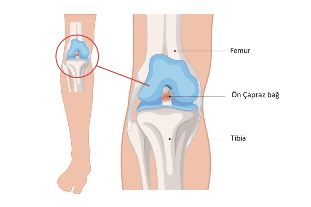
The anterior cruciate ligament (ACL) is a strong ligament located in the center of the knee joint, extending between the thigh bone (femur) and the shin bone (tibia). This ligament prevents the knee from sliding too far forward and controls unwanted movements during rotation (twisting). It can rupture, especially in athletes, during movements such as sudden changes of direction, stopping, jumping, or twisting the knee.

ACL tears are most common in sports that involve pivoting movements, such as football, basketball, and skiing. When the ligament ruptures, patients experience a “severe giving-way sensation” in the knee, along with sudden pain, swelling, and a loss of stability (the necessary tightness). Some patients report hearing a “popping or snapping” sound from their knee at the moment of the tear.

An ACL rupture seriously affects the mechanical stability of the knee. The treatment plan is determined based on the patient’s age, activity level, associated injuries (such as meniscus tears, cartilage damage, or other ligament injuries), and functional expectations. For patients who lead an active lifestyle or wish to return to sports, surgical reconstruction (rebuilding the ligament) is generally recommended.

Surgery Duration: Approximately 60–90 minutes

Type of Anesthesia: General anesthesia (in some cases, spinal anesthesia may be preferred)

Surgical Method: The procedure is performed arthroscopically (a minimally invasive "keyhole" method) by entering the knee joint.

The ruptured ligament is removed, and the ligament is reconstructed using a graft (new tissue) taken from the patient's own hamstring (back of the thigh) or patellar tendon (below the kneecap).

The graft is secured into the bone using special fixation devices (such as screws or buttons) and bone tunnels.

First day: 4–6 (This may be 2–3 if a pain pump or a nerve block is used)

First week: 3–4

After the 2nd week: 1–2

Typically 1 night in the hospital.

First 2 weeks: Use of crutches and a knee brace.

Weeks 3–6: Walking returns to normal; light exercises begin.

Weeks 6–12: Strengthening and balance (proprioception) exercises are performed.

3rd month: Light jogging may be permitted.

6th month: Return to sports activities is possible after completing return-to-sport testing.

First dressing change: Day 2

Suture removal: Day 10 (if non-dissolvable stitches are used)

Frequently Asked Questions

No. Because the mechanical stability of the knee is compromised, playing sports can lead to recurrent sprains and permanent problems, such as a meniscus tear or cartilage damage.

Non-surgical treatment may be considered for older, less active individuals who do not experience significant instability in daily life. However, reconstruction is recommended for young and active individuals.

Typically, the hamstring tendons (from the back of the thigh) or the patellar tendon (from below the kneecap) are used. This choice depends on the surgeon's preference and the patient's condition.

In this surgery, which is performed using the arthroscopic (keyhole) method, the incision scars are very small and usually fade over time.

A return to sports is generally possible around the 6th month, after the physical therapy process is complete and special tests confirm that knee function is sufficient.